Study of Worker Bee Morphology
Title: Study of Worker Bee Morphology
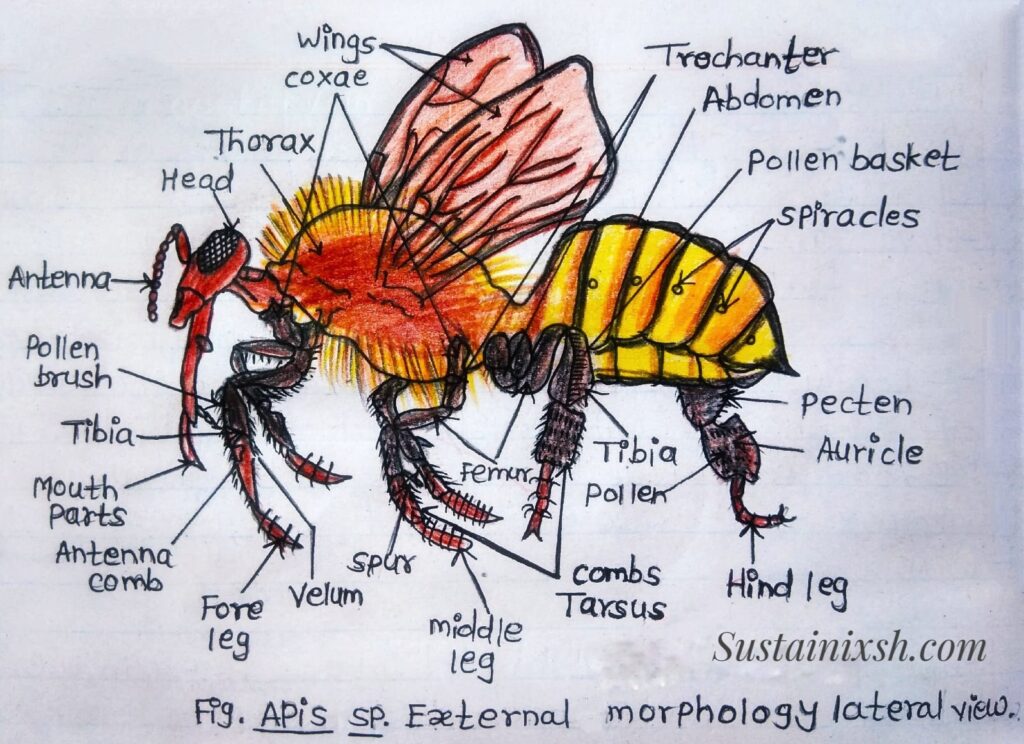
External Morphology of Worker Bee:
The worker bee is the smallest member of the colony. It is black and brown in colour, and the entire body is densely covered with hair. The body of a worker bee is divided into three regions: the Head, Thorax, and Abdomen.
• Mouth Parts: These are attached to the lower part of the head. The mouth parts are biting and Sucking type. It consists of the labrum, epipharynx, mandibles, maxillae, and labium.
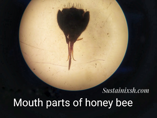
1) Labrum – Large plate structure attached to the lower margin of the clypeus.
2) Epipharynx – It lies below the labrum. Fleshy in appearance. Epipharynx is an organ of taste.
3) Mandibles – These are two in number and lie on the sides of the labrum. The mandibles of the worker are spoon-shaped, thick at the base, and narrowed through the middle. At the base of the mandibles, mandibular glands open. Mandibles are equipped with abductor and adductor muscles, which work side-to-side. Mandibles are useful to gather Pollen and mold the wax.
4) Maxillae – It lies beneath the mandibles. The lacinia is absent in the maxillae, while the maxillary palps are vestigial, and the galea is elongated and blade-like.
5) Labium – The labium shows strongly reduced Paraglossae, but the glossae are very much elongated. They are united, hairy, and form a honey-spoon called a labellum at the terminal part. The labial palps are well developed and help to make the ligula up. The apparatus is well surrounded by galeae of maxillae. At the time of nectar feeding, the labium and maxillae come together to form a sucking tube.

• Wings: The wings are small, narrow, membranous, and transparent. They lie flat over the back at rest. Wings show modified and reduced wing venation. The fore and hind wings are interlocked by hooks (hamuli), so as to work together during flight.

The wings of a worker bee are small, thin, and clear like glass. They are very light but also strong, helping the bee fly easily. When the bee is resting, its wings lie flat over its back. During flight, the wings move very fast—about 200 to 250 times every second—allowing the bee to hover, turn, and stay steady even in the wind. The front wings do most of the flying work, while the back wings help in balancing and steering. Tiny hooks called hamuli join the front and back wings together so they move as one. The wings also help the bee cool down when it gets too warm. Interestingly, scientists can tell different types of bees apart by looking at the small differences in their wing patterns.
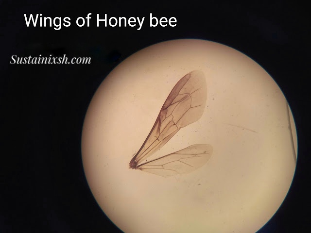
The fore and hind wings of a worker bee are intricately connected by tiny hook-like structures called hamuli. These hooks interlock the two pairs of wings, allowing them to function as a single, unified surface during flight. This interlocking mechanism provides greater stability, control, and efficiency, enabling the bee to maneuver swiftly and maintain balance while flying or hovering. The coordination of the wings through hamuli is an essential adaptation for the bee’s foraging and flight precision.

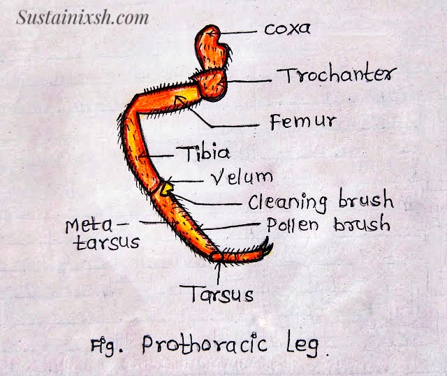
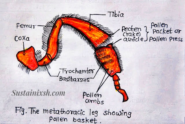
• Legs: There are three pairs of legs, i.e., prothoracic, mesothoracic & metathoracic, showing progressive increase in length from 1st to 3rd pair. Legs are densely covered with hair. Each leg consists of five parts, viz., coxa, trochanter, femur, tibia, and tarsus. The tarsus is five-jointed and ends in the claws and pulvillus. Each pair of legs is structurally modified to suit various activities/functions.

a) Prothoracic Leg – A Number of stiff bristles are present on the anterior face of the tibia distally, which form a Pollen brush. On the posterior face of the tibia, there is a movable plate-like process called velum, which fits over a circular notch in the upper part of the first tarsal segment. The velum and antenna-comb together serve as antenna cleaners.
The eye brush is present on the anterior surface of the first tarsal segment, which is used for removing pollen and other particles from the surface of the compound eyes.
b) Mesothoracic Leg – The middle leg shows the usual portion. The inner surface of the tibial segment bears a brush and a spine-like (pollen spur) at its distal end. The spurs are used to remove pollen from the pollen baskets of the hind legs and to remove wax from the wax pockets on the ventral surface of the abdomen.

c) Metathoracic Leg – Each metathoracic leg consists of a large tibia, which contains a cavity and forms the pollen basket. i.e., a depression on the outer surface of the tibia, used for storing pollens during collection. At the distal end, the tibia has a row of stiff bristles called pectins, below which is a flat plate, known as the auricle. The pectines and auricle form a pollen packet to convey packed pollens into the Pollen basket.
• Abdomen: The abdomen of a worker bee is oval and the posterior-most region of the body. It has six visible segments.
i.e., II to VII because the first segment (propodeum) is transferred to the thorax, and the remaining are reduced. Each visible segment has a large dorsal tergum and a smaller ventral sternum. The successive terga and sterna are connected by the intersegmental membrane. The posterior part of the abdomen is modified into a sting apparatus and a wax gland on the ventral surface of the abdomen.
• Sting apparatus: In the worker bee, the ovipositor changes so as to take the function of the sting, i.e., for injecting poison. The sting apparatus or poison apparatus has three components, namely, the sting, the plates, and the poison gland.

1) The Sting – The sting is a hollow organ formed by three pieces connected to a central canal (venom/poison canal). The dorsal part is the stylet sheath, and the two ventral pieces are stylets or lancets. The tips of the stylets and their sheaths have forward-directed barbs. The stylet, the sheath widens at its base into the bulb of the sting and then into a pair of twisted arms.
2) The plates – Three Pairs of plates associated with the string act as a lever. The innermost pair of oblong plates is posterior in position and represents the divided 9th sternum. Two triangular or fulcrum plates representing the reduced 8th sternum are attached to corresponding stylets. The large quadrate plates lie dorsally to the triangular plates and at the posterior angle.
3) Poison gland – The poison gland of a honey bee is a special organ found only in worker bees and the queen. It produces a venom that is used mainly for defense. When a bee stings, the poison gland releases venom through the sting (stinger) into the enemy’s skin. The venom causes pain and irritation, helping protect the hive from threats.
There are two glands –
i) Acid glands – It is an elongated, slender gland that opens into the upper end of the poison sac. It discharges an acidic secretion into the sac.
ii) Alkaline gland – It is a thick, tubular gland that opens externally below the base of the bulb.
In worker bees, the venom is strong and helps them defend the colony. However, the queen bee uses her sting and poison mainly to fight rival queens, not for defense. Although bee stings are painful for humans, the venom also has medicinal uses in small amounts, such as reducing pain and swelling in some treatments (known as apitherapy).