Study of external characters and digestive system of Starfish
Study of external characters and digestive system of Starfish
Asterias are marine & live on moist sea coasts. It is bottom dwelling hence called benthoic animal. It is a carnivorous & voracious feeder.
Systematic position:-
Phylum – Echinodermata
Sub-phylum – Eleutherozoa
Class – Asteroidea
Order – Forcipulata
Family – Asteridae
Genus– Asterias
Species – rubens
• External characters:-
1. Shape & size :-
– The body of a starfish is star-shaped, flattened in oral, aboral axis & radically symmetrical, Pentamerous arrangement. It consists of a pentagonal disc from which radiates out five, elongated, tapering arms. The axes of arms are known as radii & the regions of the Central disc between the arms are termed inter-radii. The body has an oral surface on which the mouth is situated. This surface is normally kept towards the substratum. The aboral surface is Convex & Covered with spines of various lengths. A minute opening is situated at the centre called Anus. The surface also bears madreporite at inter radii position.
2. Colour :-
Asteria is usually bright yellow, brown or orange coloured.
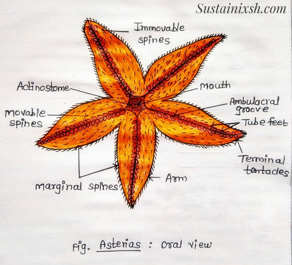
3. Oral Surface :-
The surface normally kept towards the substratum is called the oral or actinal surface. It bears a mouth, ambulacral grooves, ambulacral Spines, tube feet, eyes & Tentacles.
i) Mouth – It is a circular aperture situated at the centre of the oral surface of the central disc. It is guarded by five groups of oral spines or mouth papilla.
ii) Ambulacral groove – From the five corners of the mouth or Actinosome radiate out five narrow grooves called ambulacral grooves; which run along the middle of each arm up to its tip. Each groove shows two rows of tube feet.
iii) Ambulacral spines – Each ambulacral groove is bordered & guarded from the lateral sides by 2 or 3 rows of movable calcareous ambulacral spines. These spines are capable of closing over the groove.
iv) Tube feet or podia – Each Ambulacral groove Contains two double rows of soft, thin-walled, extensible tubular structures called tube feet, Each tube feet has a sucker disc; podium (middle) & ampulla (upper sac).
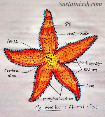
4) Aboral Surface :-
The upper convex surface is called aboral or Abactinal surface. It bears spines dermal branchiae, anus, madreporite & Pedicellariae.
i) Spines – The entire aboral surface is covered by short, stout, blunt & immovable calcareous spines or tubercles.
ii) Dermal branchiae – Those are very small, soft, delicate, hollow, finger-like membranous retractile processes present between the ossicles of the integument called dermal branchiae or gill or papula. The papula is a hollow evagination of the body wall. The dermal branchiae are respiratory in function.
iii) Anus– It is a small aperture, that lies nearly in the centre of the aboral surface.
iv) Madreporite– It is a flat, round, small but conspicuous button-like structure called madreporite. It is situated on an aboral surface eccentrically. The two rays between which madreporite is present are called bivium & three remaining rays Trivivum. The madreporite is a sieve, like a Porous place & leads to the stone canal of the water vascular system.
v) Pedicellariae – Pedicellariae are modified spines that occur in the space between the spines all over the body. These are microscopic pincer-like or jaw-like bodies. Each Pedicellariae consist of a basal Stalk which bears calcareous plates or ossicles & two joins or valves, pedicellariae are protective in function.
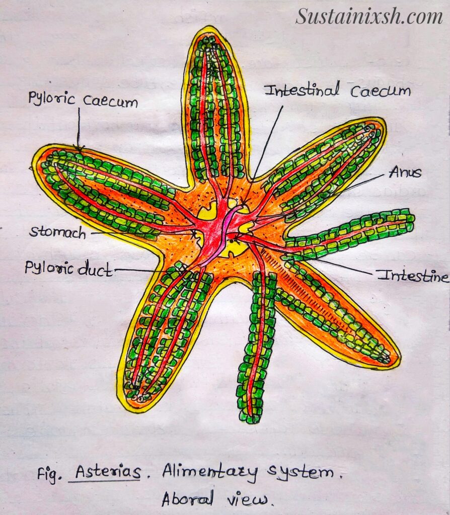
Digestive System of Starfish:-
i) Mouth – A five- rayed aperture, Present at the centre of the oral surface. It is also called Actinosome.
ii) Oesophagus – A short but wide tube connecting the mouth with the stomach.
iii) Stomach – It is divided into two by horizontal Constriction, the lower Cardiac & upper pyloric stomach. The stomach is the largest part of the alimentary canal.
a) Cardiac Stomach – It is a spacious, five-lobed sac, that occupies the greatest part of the central disc. The wall of this is thin, muscular & highly folded. It can be completely everted through mouth by Pressure of Coelomic fluid.
b) Pyloric Stomach – It is a smaller, Pentagonal sac in Communication with Cardiac stomach dorsally. Each angle of Pyloric stomach is drawn out into a duct while entering in arms & branches to form a pair of large appendages, called as pyloric caeca or hepatic caeca or gastric glands. Thus, there are five pairs of pyloric caeca, one Pair in each arm.
iv) Intestine:- A short, narrow tube runs from the Pyloric stomach to end in Anus. It gives off 2 or 3, little hollow diverticula called Intestinal or rectal caeca before opening the anus. The rectal glands are brown in colour & probably excretory in function.
v) Anus – The intestine opens on the aboral surface by a Small opening on the central disc Called Anus. It is slightly away from the centre.
vi) Digestive glands – Five pairs, long, brownish or greenish bodies, in each pyloric caecum, the hollow axis gives off laterally two series of small hollow branches, each terminating into a number of small bladder like Pouches or lobules. Digestive glands are concerned with the secretion of digestive juice containing proteases, amylases & lipase enzymes.