Human Blood smear to observe different cells
Human Blood Smear to observe different cells
We take a Microscope, glass slides, spirit lamp, sterilized lancet, a drop of blood, Giemsa stain, and Distilled water.
Preparation of Blood Smear
- Obtain 2 clean microscope slides, alcohol wipes & lancet.
- Clean a finger with an alcohol & puncture lancet.
- Place a small drop of blood at the end of the one side.
- Use another slide to make a thin blood film as per the instructions below.
- Place the second slide at a 45° angle & touch the slide with a blood drop.
- Move the spreader slide to touch the drop of blood allowing the drop to spread by capillary action along the edge of the slide.
- Immediately pull/push the slide away from the blood droplet, making a thin smear that dries quickly as you move away from the droplet.
- A perfect smear will have a ‘feathered’ edge & separated RBCs when you view it with the microscope.
● Staining the blood smear(Horizontal Staining procedure).
- Place a thoroughly dried smear on the horizontal staining rock.
- Flood smear with fixative for 10 seconds drain.
- Flood smear with fixatives for 10 seconds drain.
- Rinse the smear with DW for 1 minute.
- Air dry & examine under the microscope, using low power 1st then high power.
- Observe as many different types of blood cells as possible, and pay close attention to size, frequency & nuclear features.
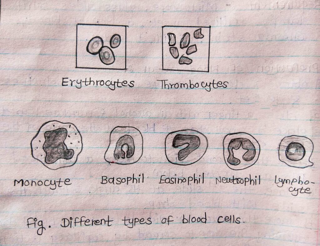
Observation:- Different types of blood cells are visible. (Neutrophils, Basophils, eosinophils & monocytes).
● Erythrocytes[RBCs]:- They are the most numerous blood cells i.e. about 4-6 millions/mm3 They are also called RBCs. In man & in all mammals. Erythrocytes are devoid of nucleus & biconcave in shape. In the other vertebrates, they have a nucleus. The red cells are rich in haemoglobin a protein able to bind oxygen. Hence, these cells are responsible for providing oxygen to tissue & partly for recovering CO2 produced as waste. In the mammalian RBC, the lack of a nucleus allows more shape for the HB & biconcave shape of these cells to raise the surface & cytoplasmic volume ratios.
● Leucocytes[WBCs]:- They are responsible for the defense of the blood 5000-7000/mm3
They are of 2 types =
(1)Granulocytes &
(2)Agranulocyte/Lymphoid cells.
The term granulocytes is due to the presence of granules in the cytoplasm of the cells. These granules have a different affinity towards neutral, acidic, or basic stains & give the cytoplasm different colors. So granulocytes are neutrophils, eosinophils, to basophils. Agranulocytes are distinguished into the lymphocytes and monocytes.
Neutrophils:- 50 to 70% They are very active in phagocytic bacteria & are present in large amounts in the pus of wounds. The nucleus is multilobed.
Eosinophils:- 2 to 4% usually bilobed nucleus coarse & strongly oxyphils granules. These cells attack parasites & phagocytes antigen-antibody complex.
Basophils:- (5 to 10%) Nucleus usually bilobed coarse basophils granules in the cytoplasm. They secrete anti-coagulant & vasodilatory substances such as histamine & serotonin.
Lymphocytes:- (20 to 40%) They are large with round nucleus & basophilic cytoplasm. They are the main constituents of the immune system which is a defense against the attack of pathogenic micro-organisms. such as viruses, bacteria, fungi & Protista.
Monocytes:- (3 to 8%) are large with a kidney-shaped nucleus, and slightly basophils.Thrombocytes/Platelets:- The main function of platelets/Thrombocytes is to stop the loss of blood from wounds(clotting). For this, they aggregated & released factor which promotes the blood coagulation. Among them, there is the serotonin which reduces the diameter of cup vessels & slows down the blood flow, and the fibrin which raps the cells from the clotting.