
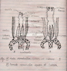
Calotes || Urinogenital (male & female) System
Urinogenital System of Calotes
A) Reproductive System of Calotes
Calotes are bisexual animals i.e., sexes in Calotes are separate. It is difficult to distinguish between the male & female from external morphological features.
i) Male Reproductive system
A pair of testes:- A pair of testes situated in the abdominal cavity that remain suspended by a special dorsal fold of the mesentery, called mesorchium. The testes are oval bodies. The testis of the right side is larger in size compared to the left side & situated slightly higher up in the abdominal cavity.
Epididymis:- From each testis’s inner surface, the epididymis proceeds posteriorly as vas deferens.
Vasa deferentia:- Two Vasa deferentia open separately by small papillae into the cloaca but before opening into the cloaca each receives the ureter of the corresponding side. So through this common duct, both urine & male gametes pass into the cloaca which is designated as the urinogenital duct.
Hemipenis:- The posterolateral side of the cloaca is provided with a pair of copulatory sacs each of which houses a hemipenis. The hemipenis is reversible. The hemipenis are grooved to conduct sperm from the cloacal cavity of the male into that of a female.
ii) Female reproductive system
A pair of ovaries:- A pair of ovaries that have a similar position as the testes. Each ovary is attached to the dorsal side by a special fold of the mesentery, called the mesovarium.
Oviduct funnel:- The anterior end of the oviduct is wide, funnel-shaped, and ciliated & is situated near the corresponding ovary below the level of the lungs.
Oviduct:- The female gonoduct is called the oviduct. The oviduct is attached to the body wall by a special fold of the peritoneum, called the broad ligament. The oviducts are not coiled but are folded and run posterior to open independently into the cloaca.
In Calotes, the fertilization is internal. Mature eggs are heavily yolked and are covered by calcareous shells.
B) Temporary mounting of scales, pecten, and hyoid apparatus of Calotes
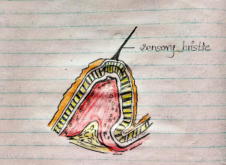
1) Scales
The skin of Calotes, like other reptiles is covered by epidermal scales. Each scale covers an epidermal & dermal scale elevation. The scales form a continuous cover over the entire body of Calotes but become thinner in the grooves between the scales.
There are two types of Scales:-
i) Larger scales
and
ii) Smaller scales lying between larger scales
Sensory structures in the form of sensory bristles are present on some large as well as small scales. These larger scales are called head shields. A few scales are called head shields. A few scales situated on the mid-dorsal line & on the borderline of the neck have become modified into larger & pointed structures. The scale cover is periodically shed by a process, called ecdysis.
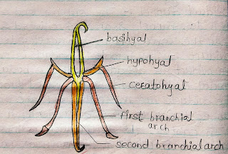
2) Hyoid Apparatus
The floor of the oral cavity is supported by a cartilaginous structure, called the hyoid apparatus. It also supports the tongue. The hyoid apparatus consists of basihyal, anterior cornua, middle cornua & posterior cornua. Basihyal adapts the body to the hyoid system. It is a median cartilaginous rod and is directed anteroposteriorly to the osentoglossus. They extend backward & outwards. Each cartilaginous rod is composed of two segments.


Curious to know in depth. Nice Blog.