
Histology of lymphoid organs
Histology of Lymphoid Organs:
Skin, Spleen, Skin Thymus, Ileum, Lymph Node, and Bone Marrow
Skin:
Type: Skin is an organ of the first line of defence against germs entering from outside the body. It is described as an anatomical barrier. It is a secondary Lymphoid organ.
Anatomy and Morphology: It is impermeable to pathogens due to the epidermis and dermis layers, as well as its low pH, which is caused by sebum secretion from the sebaceous glands.
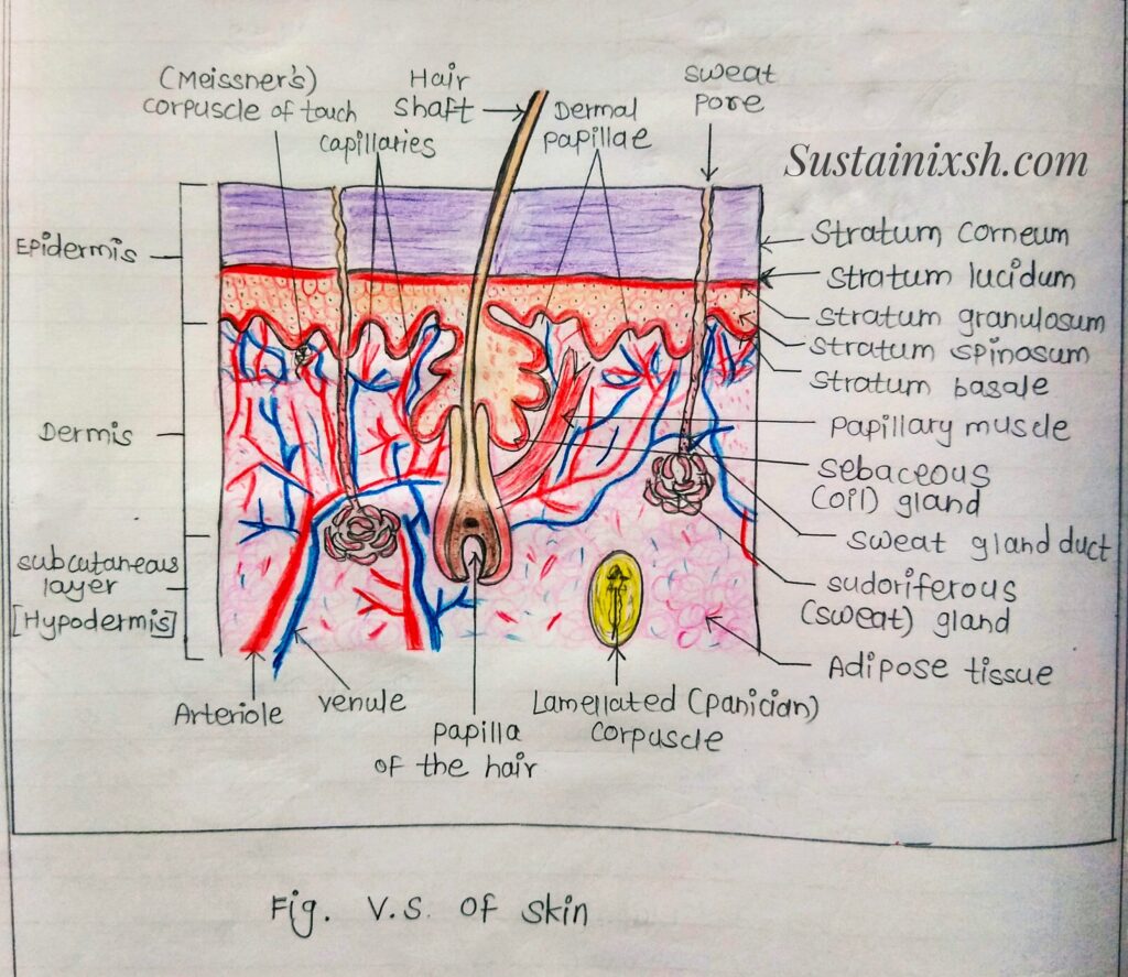
Histology: The skin is composed of epidermis and dermis.
Epidermis: It is the uppermost layer and rejuvenates after every 28 days. It consists of 5 different layers.
a) Stratum basal / germinativum: A single row of cells attaches to the dermis and youngest cell.
b) Stratum spinosum /prickle: Made up of bundles of Proteins that resist tension.
c) Stratum granulosum: Layer of flattened keratinocytes produces keratin.
d) Stratum lucidum / clear: Layer present only on the palm and soles.
e) Stratum Corneum: Horny cornified superficial layer.
● Epidermis is made up of variety of cell types:
Keratinocytes: Cells containing keratin, these are stratified Squamous epithelial cells. They arise from basal layer calls.
Non-keratinocytes: Includes melanocytes, lymphocytes and Langerhans’ cells. Melanocytes synthesise and store melanin. Langerhan’s cells serves as antigen presenting cells.
Dermis: Below the epidermis, dermis is present. Irregularly arranged and filled mostly with connective tissue. It contains two types of cells:
i) Permanent cells: like arrector pilli muscles, Vessels and nerves.
ii) Migratory cells – Lymphocytes, mast cells, NK cells.
Spleen:
Type: secondary Lymphoid organ.
Location: Situated on the left side of abdominal cavity below Pancreas.
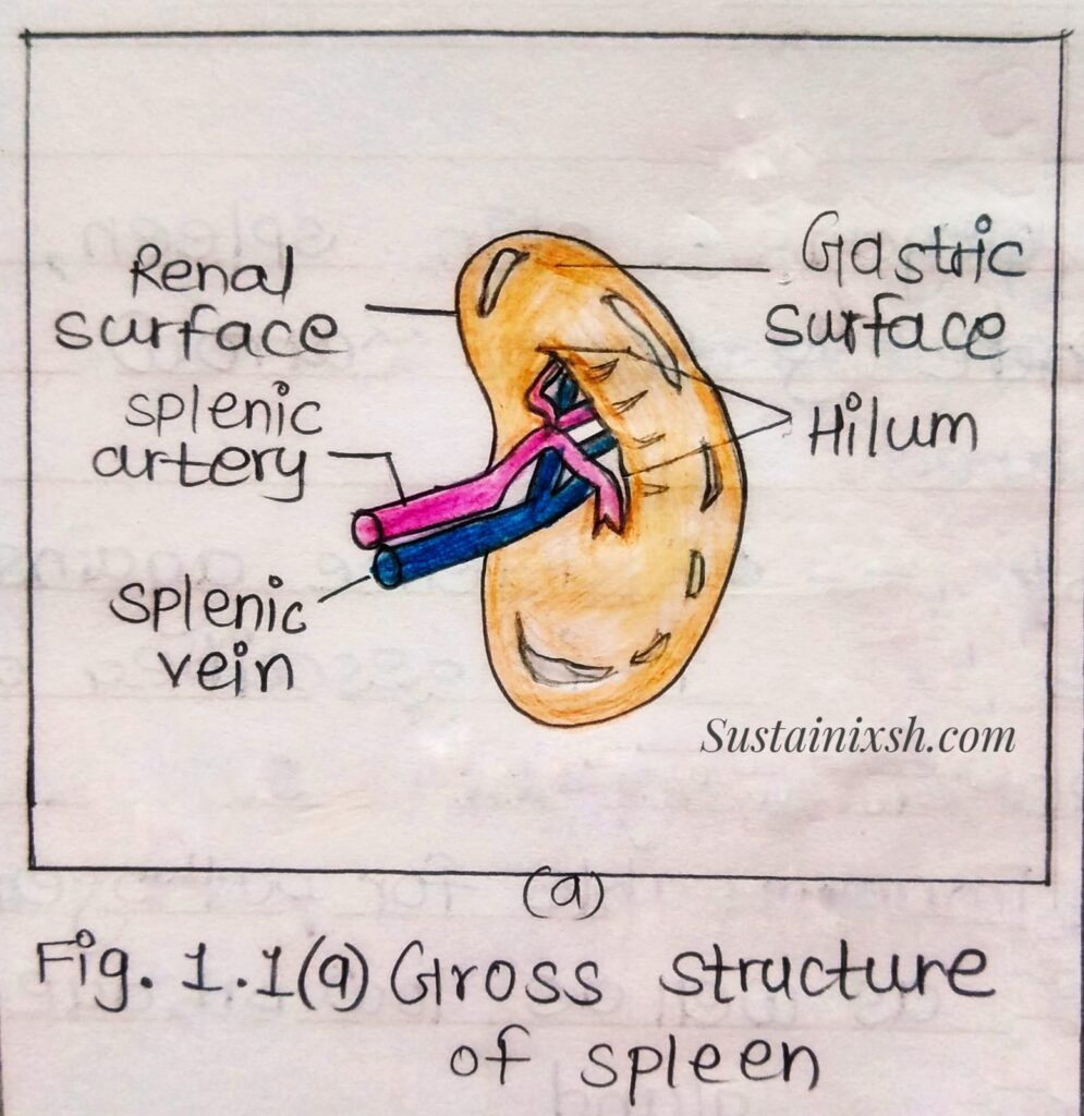
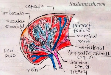
Morphology: Bean-shaped, red coloured encapsulated organ. It is the largest lymphoid organ with a depression called hilum. Entire spleen is covered by serosa membrane peritoneum except at hilum.
Histology:
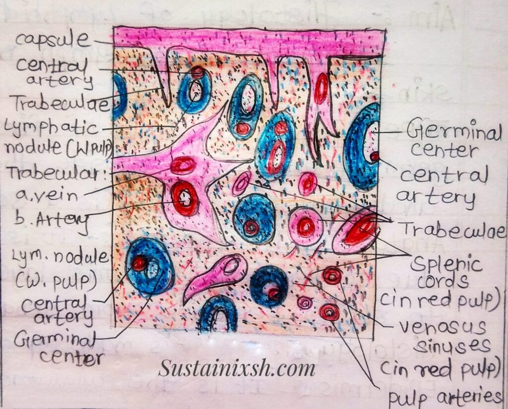
- Beneath the Peritoneum, elastic fibrous connective tissue and smooth muscles collectively forms the capsule.
- From the interior of the capsule, many trabaculae arises.
- From the hilum spleenic vessels (artery and vein) make the entrance and exit.
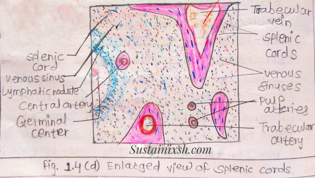
- The parenchyma contains the (a) White pulp and (b) Red pulp.
a) White pulp: surrounds branches of the spleenic vessels (artery) forming a periarteriole lymphoid sheath (PALS) mainly consisting of T-lymphocytes. Primary lymphoid follicles are attached. To PALS rich in B cells. Secondary lymphoid follicles present a deep interior and contain a germinal centre along B-cells, which develops when antigenic challenges to primary follicles are seen.
Red pulp: It consists of a network of sinusoid with macrophages red blood cells, and a few lymphocytes. It is a site for destruction of old and defective RBCs.
Functions:
- Spleen macrophages destroy blood born pathogen via phagocytosis.
- Within the red pulp, removal of ruptured defective blood cells and Platelets; storage of Platelets (1/3rd).
- Haematopoiesis during foetal life.
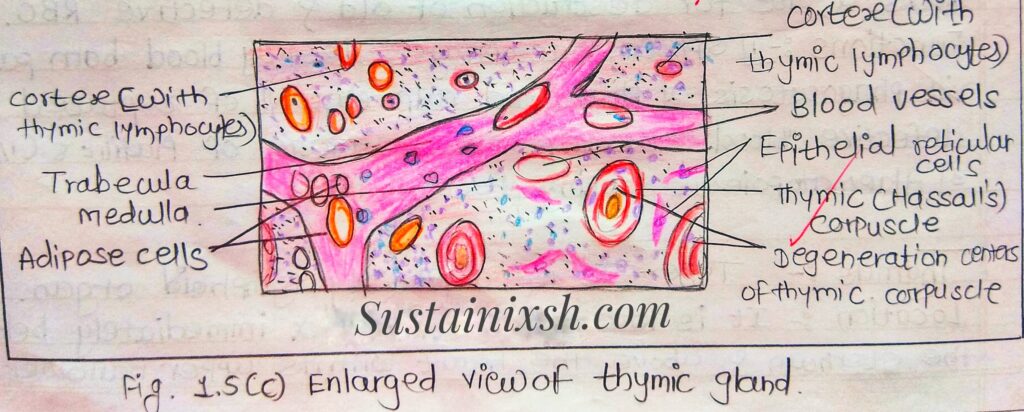
Thymus:
It is a primary lymphoid organ.
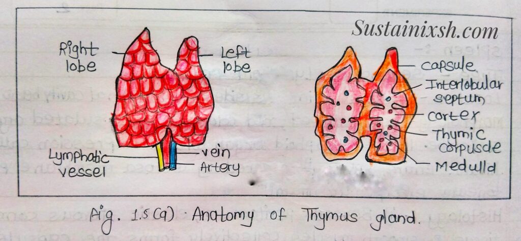
Location: It is located in the thorax immediately below the sternum and above the heart, with its upper narrower part extended into the neck, Extends between thyroid gland fourth costal cartilage within the superior mediasternum and the anterior part of inferior mediasternum.
Anatomy and Morphology: In the early stage of development it is entirely a epithelial structure derived from the third and fourth pharyngeal pouches.
The epithelial structure gradually grows and descends to the thorax, losing the connection with the pharyngeal pouches. Progenitor lymphocytes of haematopoietic cells migrate from bone marrow to the thymus through the bloodstream for differentiation and maturation into immunocompetant lymphocytes.
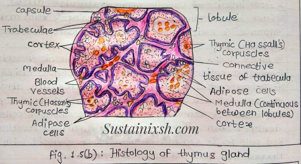
Histology: The fully developed thymus is encapsulated by connective tissue and becomes a bilobed structure. The number of lobes in thymus is varies in different animals. The Connective tissues of capsule penetrates deep into the lobes dividing each lobe incompletely into many lobes. The strands of Connective tissue in between the lobes are known as “trabacules”.
Each lobules in thymus organized into an outer cortex and inner medulla. Cortex or peripheral part of each lobule is diversely populated with differentiated but immature ‘T’ cells or thymocytes, and the medulla or central portion of lobule is Packed with loosely arranged mature lymphocytes. The hormone thymosin, secreted by thymus epithelial cells, might be helping in the differentiation and maturation of the T-lymphocytes.
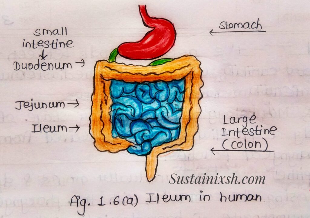
Ileum:
Type- It is a type of secondary lymphoid organ.
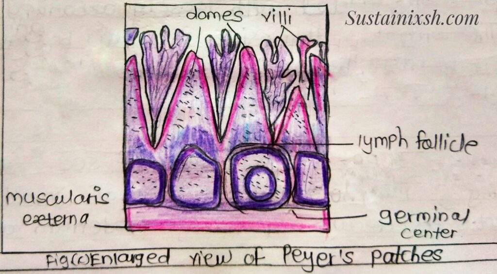
Anatomy and Morphology- last part of small intestine which connects to caecum separately duodenum and jejunum by ileocecal valve(ICV). Ileum along with jejunum, is suspended inside. mesentry lymphatic vessels and nerve fibres. Ileum has abundant payer’s patches unencapsulated lymphoid nodule that contains large number of lymphocytes and cells of immune system.
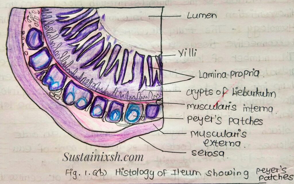
Histology: It is made up of four layers, which make up the wall of the ileum: that are mucosa membrane, the Submucosa, the external muscularis layer and the serosa.
i) Mucosa membrane: Formed by three layers.
(a) single layer of tall cells lining the lumen of organ. Epithelium the innermost layer has 5 distinct type of cells introcytes with microvilli, goblet cells, Paneth cells (release antimicrobial substance like and- defensive and lysozymes) most common in terminal part, microfold cells(take up and transport Ag from lumen to lymphatic cells of lamina Propria) and entero endocrine, cells hormone).
(b) Lamina propria – Composed of loose connective tissue large aggregate of lymphoid tissue called payer’s patches.
(c) Thin layer of smooth muscle called muscularis mucosa.
ii) Submucosa: Formed by dense irregular connective tissue caries large blood vessels and nervous component Called submucosal plexus, which is part of enteric nervous system.
iii) External muscular layer: Formed by a layers of Smooth muscles arranged in circular bundles in inner layer and longitudinal bundles in outer layer.
iv) Serosa: Composed of mesothelium, a single layer of flat cells with varying quantities of underlying connective and adipose tissues.
Functions:
- Enzymatic digestion of nutrients.
- Absorption of Vit B 12, fats and bile salts.
- Immunological Functions.
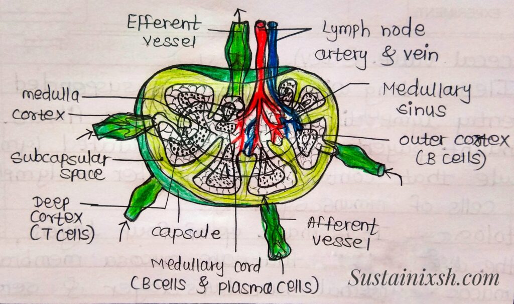
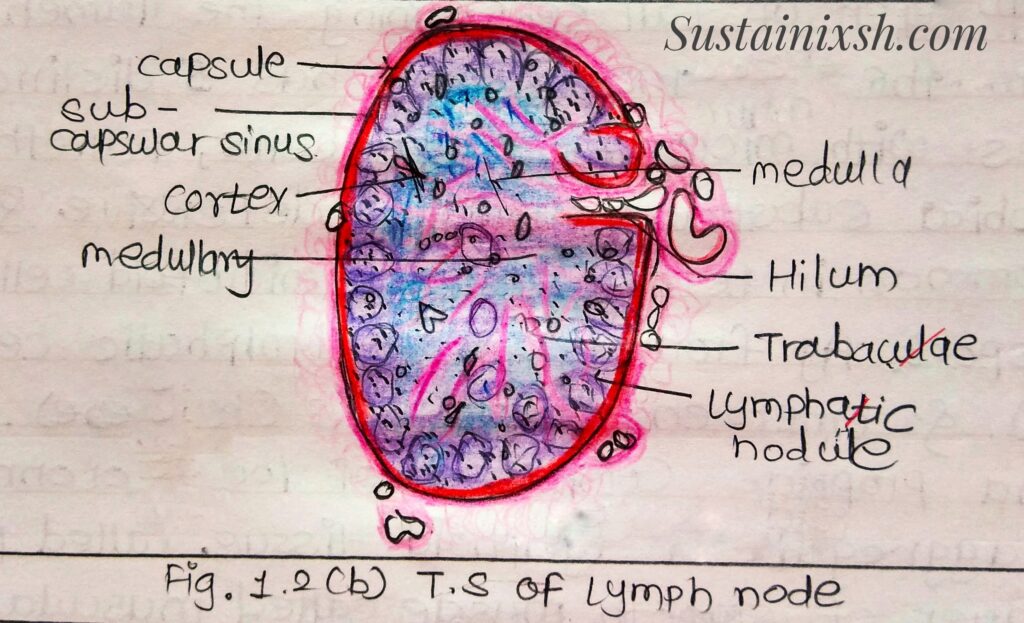
Lymph Node:
Type: It is a secondary lymphoid organ.
Anatomy and Morphology:
Location: Found all over the body including neck, armpits, brain, around the gut and between the lungs.
Structure: Lymph nodes are fully commited to regulating an immune response. They are encapsulated bean shaped structure that includes network of stromal cells. (ie. supportive tissue) packed with lymphocytes, macrophages and dendritic cells.
Connected to both blood vessels and lymphatic vessels, lymph nodes are the first organised lymphoid Structure to encounters antigens that enter tissue space.
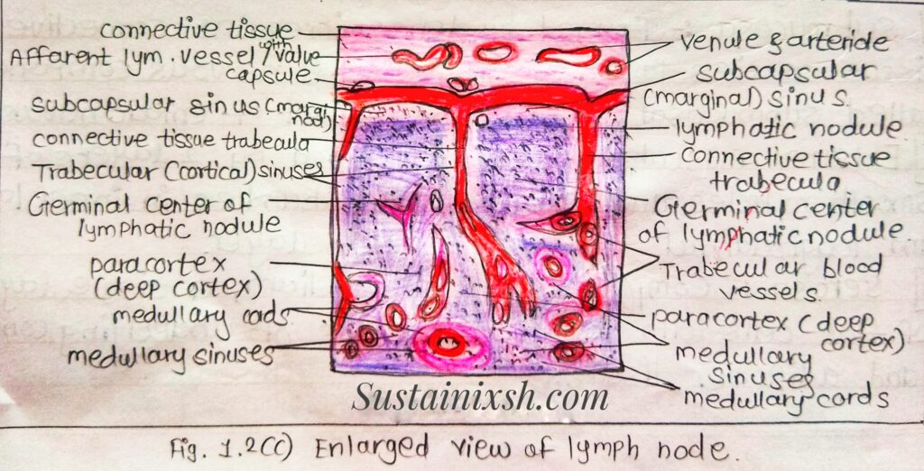
Histology: It has three concentric regions: cortex, paracortex and medulla.
Cortex: It is the outermost layer. It contains lymphocytes (mostly B cells), macrophages, follicular, dendritic cells arranged in follicles.
Para Cortex: It is present beneath the cortex. Para cortex is populated largely by T. lymphocytes but also contains dendritic cells. Dendritic cells are migrated from surrounding tissue.
Medulla: It is the innermost layer. It is the site where lymphocyte unit the lymph node through the outgoing lymphocytes.
Functions: Lymph node Provides idea microenvironment for encounter between antigens and lymphocytes, and is protective. Organised cellular and humoral immune response.
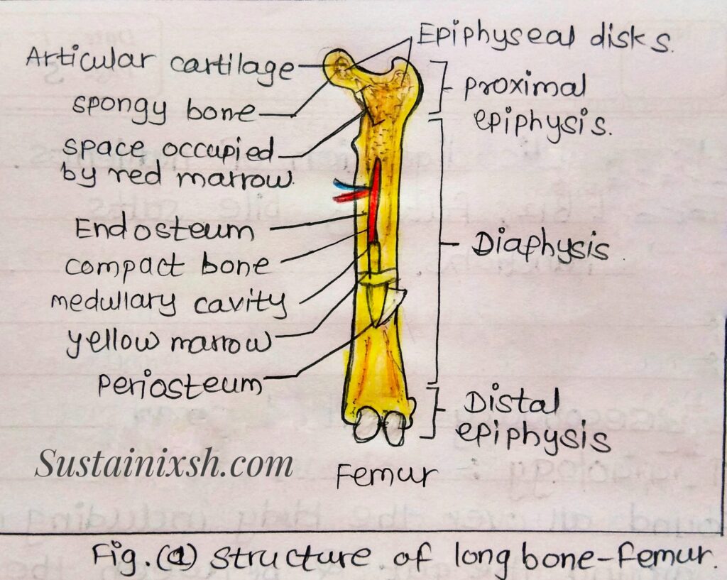
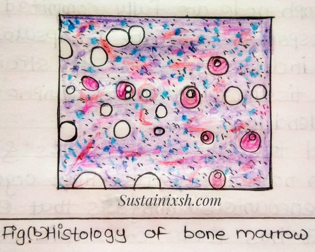
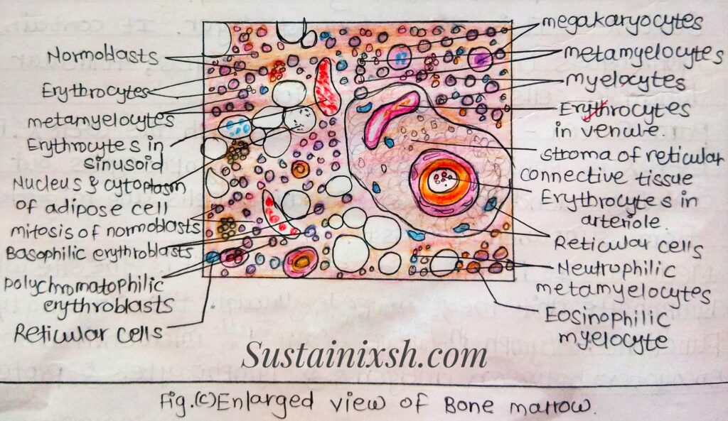
Bone Marrow:
Type: It is a primary lymphoid organ.
Location: It occurs in long bones like femur, tibia, Fibula, etc.
Structure: It is soft, spongy, gelatinous tissue. It is highly cellular composed of haematopoietic cells in various stages of development, supportive reticular connective tissue and sinusoid complex, capillaries. It Consists of delicate reticular connective tissue mesh with various cells in between.
Cell type: Conspicuous in the marrow are large adipose cells with large vacuoles, and small peripheral cytoplasm is seen. Other cells include megakaryocytes, myelocytes, granulocytes and many erythrocytes. Bone marrow smear- pleuripotent stem cells are capable of differentiating into stem cells for different haematopoietic lineages on the basis of colonies in the spleen, thus are called colony-forming units or CFU. In adults greatest concentration is found in bone marrow.


Lymphoid organs are really important for life. They works like our body organs guards